Nontuberculous Mycobacterial Lung Disease Caused by Mycobacterium shinjukuense: The First Reported Case in Korea
Article information
Abstract
Mycobacterium shinjukuense is a novel species of nontuberculous mycobacteria (NTM) that was first reported in Japan in 2011. It is a slow-growing NTM pathogen that can cause chronic pulmonary infections. There are only a few reported cases of M. shinjukuense infections, all of which are from Japan. We reported a case of chronic lung disease caused by M. shinjukuense. The organism was identified by 16S rRNA, rpoB, and hsp65 gene sequencing. To the best of our knowledge, this was the first confirmed case of lung disease caused by M. shinjukuense outside of Japan.
Introduction
The class of nontuberculous mycobacteria (NTM) is generally comprised of mycobacteria other than Mycobacterium tuberculosis complex and Mycobacterium leprae. The prevalence of lung diseases caused by NTM is increasing worldwide, including in Japan and South Korea1234. NTM are ubiquitous in the environment. Therefore, it is crucial to isolate and identify the causative organisms for diagnosis. There are some specific diagnostic criteria that have already been proposed5.
Mycobacterium shinjukuense, a novel slow-growing NTM pathogen, was introduced as a new species in 20116. There are only a few reported cases of M. shinjukuense that met the diagnostic criteria for NTM lung disease6789. All of the prior cases were reported from Japan. We report a case of NTM lung disease caused by M. shinjukuense, which was identified using 16S rRNA, rpoB, and hsp65 gene sequencing. To the best of our knowledge, this is the first case of M. shinjukuense lung disease in Korea.
Case Report
A 56-year-old Korean woman with a history of pulmonary tuberculosis (10 years prior to presentation) was referred to our hospital with a mild, but persistent productive cough for three months. Otherwise, the patient was a healthy, nonsmoker. She has not lived in Japan.
A chest radiography revealed subtle crowding of the bronchovascular bundle in both lower lung zones. A computed tomography scan of her chest revealed several nodules, micro-nodules, and bronchiectasis with lung atelectasis in the right middle lobe and the lingular division of the left upper lobe (Figure 1). There was no organic growth on the initial microbiological study of three sputum specimens. However, cultures of two bronchial washing specimens demonstrated NTM growth.
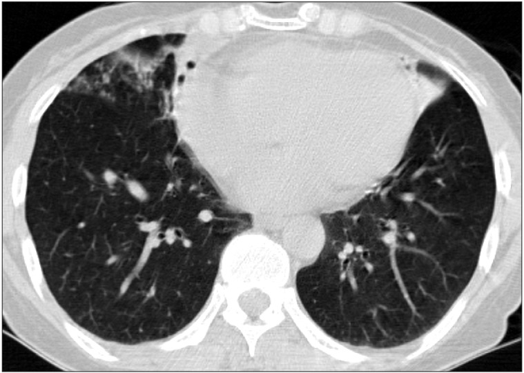
A 57-year-old female with a chronic cough productive with sputum. Axial computed tomography (CT) imaging shows several nodules, micro-nodules, and bronchiectasis with lung atelectasis in the right middle lobe. It also reveals bronchiectasis with lung atelectasis in the lingular division of the left upper lobe. These are typical CT findings of the nodular bronchiectatic form commonly seen in nontuberculous mycobacterial disease.
Initially, a polymerase chain reaction (PCR)-reverse blot hybridization assay method based on rpoB gene (REBA Myco-ID; M&D Inc., Wonju, Korea) was used to identify the species. However, this method was unsuccessful, because there were no available reference sequences of the organism1011. Next, we performed sequencing analysis of the nearly complete 16S rRNA gene sequence and partial sequences of the rpoB and hsp65121314. The 16S rRNA, rpoB, and hsp65 sequences were 100% identical to those of the M. shinjukuense type strain GTC 2738 (GenBank accession Nos. NR112623, AB268504 and AB268505, respectively). Phylogenetic analysis was performed the rpoB sequences from the isolated SMC-shi-001 strain, and sequences of closely related species within the slow growing mycobacteria class. It revealed that the isolated strain belongs to M. shinjukuense (Figure 2). The GenBank accession numbers of 14 species' sequences were compared to that of SMC-shi-001. The sequences were obtained from the GenBank sequence database.
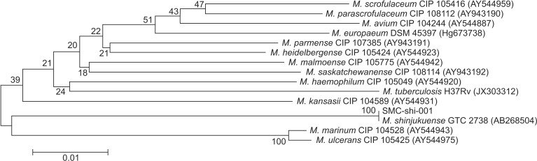
The phylogenetic position of isolated SMC-shi-001 strain from the patient in this report and other species belonging to the slow growing mycobacteria, based on the rpoB sequence. This tree is constructed using the neighbor-joining method. The percentages indicated at nodes represent bootstrap levels supported by 1,000 re-sampled datasets. Scale bars indicate evolutionary distance in base substitutions per site. M., Mycobacterium.
After the isolate was identified, we planned to start antibiotics because of persistent productive cough and progression of multiple nodules on the chest radiography during 6 months of follow-up. However, the patient was reluctant to receive long-term antibiotic therapy and refused further follow-up.
Discussion
NTM species are well known for causing chronic pulmonary disease. Some of the most commonly isolated species are the Mycobacterium avium complex, Mycobacterium abscessus complex, and Mycobacterium kansasii5. Other NTM species have also been implicated in lung disease. More than 150 NTM species have been discovered, and the number is still increasing as mycobacteriology methods continue to improve15. M. shinjukuense is a novel species of Mycobacterium that was first reported by Saito et al.6 in Japan in 2011. M. shinjukuense was described a slow growing, non-chromogenic Mycobacterium species. Compared to other mycobacteria, M. shinjukuense has unique gene sequences. Although it is phylogenetically related to M. tuberculosis, Mycobacterium ulcerans, and Mycobacterium marinum, it demonstrated <70% reassociation in DNA-DNA hybridization6.
It is essential to accurately identify isolated NTM species in order to select an appropriate antimicrobial regimen. However, commercial kit for the identification of NTM species cannot identify M. shinjukuense. These kits use techniques such as PCR-restriction fragment length of polymorphism analysis or REBA Myco-ID based on the rpoB gene. However, they cannot detect newly defined or infrequent species, such as M. shinjukuense. In this case, M. shinjukuense was not initially identified with a commercially available kit, but was with gene sequencing of the 16S rRNA, rpoB and hsp65 genes. To the best of our knowledge, this is the first case of lung disease caused by M. shinjukuense occurring outside of Japan.
According to previous reports, M. shinjukuense has been isolated from sputum and bronchial lavage fluid from Japanese patients with bronchiectasis and/or cavitary lesions6789. None of these patients was immunocompromised. There are only two case reports that address the antibiotic treatment of M. shinjukuense. In one case, the clinical efficacy of combination therapy including clarithromycin, rifampin, and ethambutol was addressed7. In another, the efficacy of the standard anti-tuberculosis drugs including isoniazid, rifampin, and ethambutol was explored89. We could not evaluate treatment efficacy; however, because the patient was reluctant to undergo long-term antibiotic treatment.
In conclusion, this is the first report of NTM lung disease caused by M. shinjukuense outside of Japan. M. shinjukuense should be considered as a potential causative pathogen in cases of NTM lung disease. Its diagnosis can be verified by performing multilocus sequence analysis of rpoB, hsp65, and 16S rRNA fragments from clinical isolates.
Acknowledgements
This work was supported by a grant of the Korean Health Technology R&D Project, Ministry for Health & Welfare, Republic of Korea (A120647) and by the Basic Science Research Program through the National Research Foundation of Korea (NRF) funded by the Ministry of Education (2013R1A1A2060552).
Notes
Conflicts of Interest: No potential conflict of interest relevant to this article was reported.